George Crawford, MD
Ventral Hernia Repair - Medium Hernia with Mesh
![](https://d15k2d11r6t6rl.cloudfront.net/pub/bfra/jdk0tdba/fbw/yh1/q2x/Screenshot%202025-02-06%20102705.png)
Procedure Overview
Ventral hernias can be categorized as incisional, umbilical, or epigastric.
Medium ventral hernias can be repaired by excising the hernia sac and then using a mesh to close the fascia defect.
INDICATIONS:
- Ventral hernia repair may be indicated to prevent or address symptoms related to the hernia such as pain, acute incarceration, enlargement, and skin problems.
- The most common sites for ventral hernias are the navel (an umbilical hernia) and the site of a previous abdominal procedure (incisional hernia).
![](https://d15k2d11r6t6rl.cloudfront.net/pub/bfra/jdk0tdba/1sx/os5/djy/orthopedic-surgery-orthopedics-polio-7817270.jpg)
Relevant Anatomy
Click to interact with a BioDigital Human animated 3D anatomy, disease states, and procedure tour.
Pre-operative Patient Care
Typical recommendations for pre-operative care may include, but are not limited to, any of the following:
- CT Scan with PO and IV contrast.
- Cardiopulmonary evaluation as needed.
- Anesthesiology consultation as needed.
- Nothing by mouth for 6 hours before surgery.
- Prophylactic antibiotics for patients with comorbidities such as heart disease, diabetes, previous history of colon or bowel surgery.
- BMI greater than 40 and smoking are relative contraindications.
Prep & Patient Positioning
Common patient positioning:
- A supine position with the arms tucked at the patient's sides, for patients with midline hernias.
- A lateral decubitus or modified lateral decubitus position, for hernias of the flanks or posterior abdominal wall.
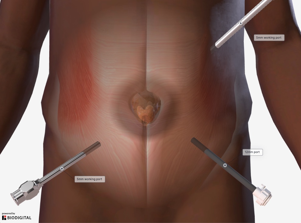
Common port placement
- The location of initial abdominal access (primary port placement) for laparoscopic ventral hernia repair is typically as far from the hernia defect and prior laparotomy incisions as possible.
- A Veress needle, open Hasson technique, or optical trocar entry may all be used for primary port placement. The specific technique used should be primarily based on the surgeon’s experience and outcomes with the technique and take into consideration the patient’s surgical history and anatomy.
- Secondary port placement is generally performed under direct vision and placed as lateral from the hernia defect as possible to allow the surgeon to work in an ergonomically favorable position.
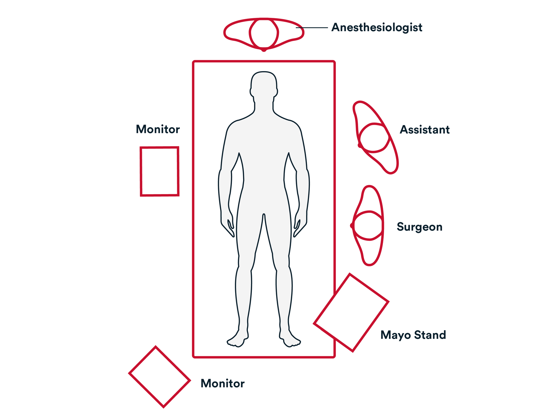
![](https://d15k2d11r6t6rl.cloudfront.net/pub/bfra/jdk0tdba/32e/yr5/8h1/Medium_Hernia_Port_Placement.jpg)
![](https://d15k2d11r6t6rl.cloudfront.net/pub/bfra/jdk0tdba/rsm/rm6/cbc/Hernia%20Ventral%20Medium.png)
Operative Steps
![](https://d15k2d11r6t6rl.cloudfront.net/pub/bfra/jdk0tdba/31r/qb7/ojv/1_1.png)
Access
![](https://d15k2d11r6t6rl.cloudfront.net/pub/bfra/jdk0tdba/31r/qb7/ojv/1_1.png)
Access
![](https://d15k2d11r6t6rl.cloudfront.net/pub/bfra/jdk0tdba/gi1/j1q/o12/Arrow_Down.png)
- Access is generally obtained through an incision in the left upper quadrant. Usually, a 5 mm trocar is used to enter the abdomen if laparoscopic assistance is needed.
- Once visualized and safe, a left lower quadrant 12 mm trocar and a right lower quadrant 5 mm trocar are placed under visualization.
- All abdominal wall adhesions are taken down using blunt dissection, sharp dissection, or an energy device. This is done to remove adhesions to visualize the ventral hernia defect.
- Any intra-abdominal content, such as small bowel, is reduced from the hernia.
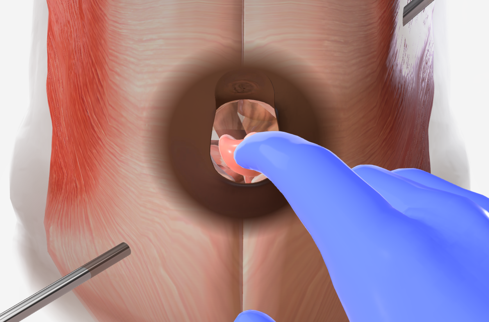
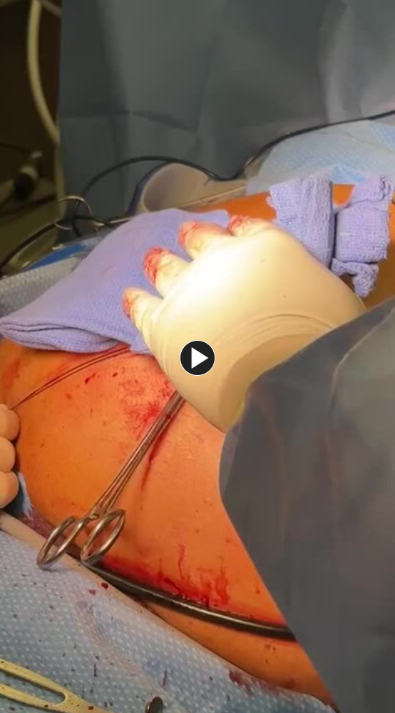
- An incision is made over the ventral hernia. The subcutaneous tissue is dissected to expose the hernia sack.
![](https://d15k2d11r6t6rl.cloudfront.net/pub/bfra/jdk0tdba/pc5/csr/ccj/2_1.png)
Repair
![](https://d15k2d11r6t6rl.cloudfront.net/pub/bfra/jdk0tdba/gi1/j1q/o12/Arrow_Down.png)
![](https://d15k2d11r6t6rl.cloudfront.net/pub/bfra/jdk0tdba/pc5/csr/ccj/2_1.png)
Repair
- The hernia sac is dissected out bluntly and exposed.
- Once completely exposed, the hernia sac is excised at the base of the fascial defect. The fascia is exposed circumferentially around the defect.
- A mesh, such as the PROCEED™ Ventral Patch is placed through the fascia
- It is secured to the facia in all four quadrants, commonly using a size 0-Prolene suture and a suture passer. Any gaps in the repair from the fascia to the abdominal wall are closed using an absorbable laparoscopic fixation device such as ETHICON SECURESTRAP™.
- A suture, such as Coated Vicryl™ Plus Antibacterial suture, Prolene™ Polypropylene, or Ethibond EXCEL™ Polyester can be used to re-approximate the facia over the mesh if possible.
- If this is an umbilical hernia, and the umbilicus has been mobilized away from the facia, it may be reapproximated, using synthetic absorbable suture, such as a size 0-Coated Vicryl™ Plus Antibacterial suture.
![](https://d15k2d11r6t6rl.cloudfront.net/pub/bfra/jdk0tdba/y0u/ye2/246/3_1.png)
Closure
![](https://d15k2d11r6t6rl.cloudfront.net/pub/bfra/jdk0tdba/gi1/j1q/o12/Arrow_Down.png)
![](https://d15k2d11r6t6rl.cloudfront.net/pub/bfra/jdk0tdba/y0u/ye2/246/3_1.png)
Closure
- The trocars are removed. The wounds are closed using synthetic absorbable suture, such as Monocryl™ Plus Antibacterial suture, and a topical skin adhesive such as DERMABOND™ PRINEO™ Skin Closure System or any appropriate dressing.
Post-operative Patient Care
Typical recommendations for post-operative care may include, but are not limited to, any of the following:
- The patient is out of bed, ambulating, with appropriate pain control.
- Diet is advanced as tolerated, with plans for discharge on postoperative day 0 for routine umbilical hernia.
- Lift nothing heavier than 25 lbs. for two weeks.
- Resumption of normal activity, minus lifting restrictions, occurs within 1 day following the procedure.
- Discharge instructions consist of pain management, instructions on the future signs and symptoms indicating potential complications (see below), and an office appointment in 1 weeks’ time.
Potential complications may include but are not limited to, any of the following:
- Nausea, vomiting, or severe coughing spells.
- Fever greater than 101.5 degrees.
- Redness or Erythema.
Additional resources
![](https://d15k2d11r6t6rl.cloudfront.net/pub/bfra/jdk0tdba/kcl/frt/pa3/woman-in-blue-ppe-wearing-blue-face-mask-4586994.jpg)
Access our on-demand Hernia video library
Looking for more? Explore procedural videos and webinars from global experts
![](https://d15k2d11r6t6rl.cloudfront.net/pub/bfra/jdk0tdba/iez/lsu/vap/HERNIAcademy%20Featured%20Program%20Image.jpg)